ucla and U of T researchers have teamed up to discover the chemical processes that occur when seals sleep, and their discoveries have been published in The Journal of Neuroscience.
Fur seals have a fascinating sleep pattern: on land, electroencephalogram (eeg) activity is observed bilaterally. However, in water, they exhibit asymmetric slow-wave sleep (asws) brain activity, which is similar to that of toothed whales. The eeg activity of one hemisphere is low-frequency and high-voltage while the other is high-frequency and low-voltage. Interestingly, the foreflipper on the opposite side of the active part of the brain moves to maintain body orientation; the eye on this side is also open.

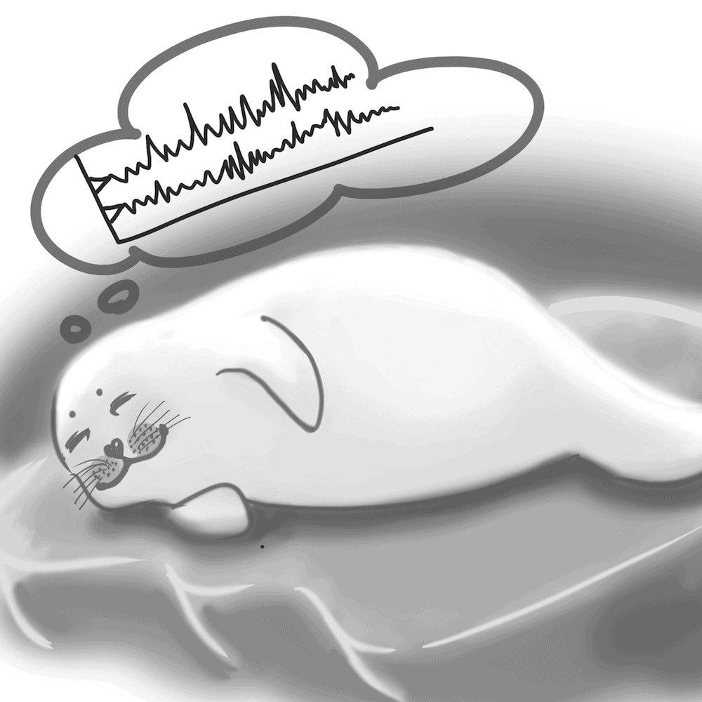
NANCY JI/THE VARSITY
Knowledge of the neurotransmitters involved in sleep biology mostly comes from species with bilaterally symmetrical eeg patterns. Researchers have already discovered that when fur seals sleep on land, levels of acetylcholine are at thier highest during wakefulness and lowest while sleeping. When fur seals sleep in water, there is greater acetylcholine-dependent activity in the hemisphere that demonstrates the lower voltage, higher frequency eeg patterns.
The Journal of Neuroscience paper studied the neurotransmitter studied was serotonin. Serotonin has been known to play a major role in inducing and maintaining sleep. For instance, when there is a depletion of serotonin one suffers from insomnia. The study of rat brain slices has shown that serotonin may target sleep-active neurons in the pre-optic area of the brain.
In order to study the role of this chemical during sleep in fur seals, scientists implanted electrodes symmetrically in both hemispheres of four juvenile fur seals from the Commander Islands in Russia. These seals were given one year to adapt to their surroundings and new environment in captivity so that the research findings would be as accurate as possible. The scientists used a procedure called microdialysis to sample the serotonin from the brains of these fur seals and to take a serotonin assay in order to detect the amount of serotonin at various stages of sleep.
When fur seals slept on land, serotonin was expressed maximally in both hemispheres during non-rem sleep and minimally during rem sleep. During non-rem sleep on land, serotonin release was not significantly different between each hemisphere. This is in contrast to acetylcholine release,
which is elevated in the waking state, while
serotonin is elevated in the sleeping state.
Serotonin has previously been studied in relation to its effect on mood in humans. For instance, selective serotonin reuptake inhibitors (ssris) have been used to modulate mood. It
has also been shown that the effect of serotonin on mood may be indirect rather than direct, as it takes weeks until there are observable changes in mood. Hence, the indirect link may be due to slow neurological changes rather than an immediate change in serotonin level.
Some scientists have postulated that one factor driving the switch between bilateral or unilateral hemispheric slow-wave sleep is the corpus callosum, the network of nerve fibres in the brain that connects the left and right hemispheres. Interestingly, toothed cetaceans, such as dolphins, exhibit only unilateral hemispheric slow-wave brain activity never the bilateral eeg brain activity of land mammals. The size of the corpus callosum of these cetaceans is much smaller than in mammals. However, scientists have not yet found a direct correlation between size of the corpus callosum and type of slow-wave sleep.
This research study concludes that there are many factors involving the onset of sleep and various types neurotransmitters, such as serotonin and acetylcholine, that are involved. Future studies will include the analysis of the effects of histamine, hypocretin cells, and norepinephrine on sleep.