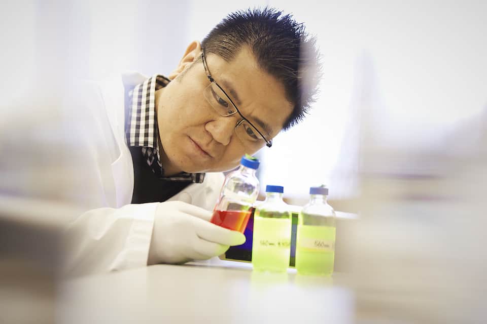
Dr. Warren Chan of UofT’s Institute of Biomaterials and Biomedical Engineering, along with PhD student Dylan Glancy and postdoctoral fellow Seiichi Ohta, have managed to “shape-shift” nanoparticles in their most recent study.
Extensive research on nanoparticles has demonstrated a strong potential for the small objects to contribute to the highly localized delivery of drugs in the treatment of cancer. Some researchers speculate that within half a century, we will look back at the practice of injecting freely transported drugs into our circulatory system as a crude convention of the past. While having Aspirin molecules freely floating in your blood is usually benign, one cannot say the same for commonly used chemotherapy drugs.
Nanoparticle design has two primary issues: first finding, and then interacting with the correct cell and not its neighbours.
Nanoparticles must survive the trip through the blood stream, evade the body’s immune system, and, in the case of cancer, penetrate deep into dense tumor tissue and into individual cells.
Once there, the right cell is targeted through the use of “lock and key” arrangements, where the nanoparticle snugly latches on to a specific molecule, unique to the affected cells.
Unlike most nanoparticles, those developed by Dr. Chan and his associates have the ability to switch shapes, from a rod to a sphere. “Rod-shaped nanoparticles for example are good at penetrating tumors, while spherically shaped ones are absorbed by cells more easily.” explained Glancy. “Most of the currently existing dynamic nanoparticles might permanently lose an outer layer or increase in size under different conditions, but they don’t change shape, and they definitely don’t do it reversibly.”
This reversibility will be helpful in the removal of the nanoparticles from out of the tumor and into the blood stream after they have done their job.
This shape change is brought about by the use of flexible adhesive strands of DNA known as “biological Velcro.” There are two core nanoparticles of different sizes strung together by DNA, with the larger core being “orbited” by smaller “satellite” particles. All of the particles have sticky DNA threads attached to their surface.
Introduction of loose DNA can help link the DNA on the satellites simultaneously to both core particles, and not just one. The introduction of additional competitive loose DNA helps break the linkage between the satellite particles and the larger core by removing the original linker DNA. This graceful displacement, described by Glancy as “DNA origami,” results in an elongation of the resultant structure from a sphere to a rod and vice versa.
The beauty of using DNA is that it can be designed to simultaneously adhere to specific DNA molecules expressed by faulty cancer cells. Alternatively, the particles can additionally be decorated with proteins that are known to interact with other proteins found on the surface of cancer cells, due to the large surface area of the nanoparticles. Examples of “lock and key” pairs suggested by Dr. Chan include the popular antibiotic Herceptin and HER2 receptors, as well as folic acid and folate receptors, both of which are receptors commonly displayed in breast cancer cells.
“This can be very useful in targeting cancer cells that have metastasized, which are extremely hard to localize through surgery for example” added Glancy, “and since they are usually found closer to blood vessels, they can fortunately be easily reached by the blood transported nanoparticles.”
Once the nanoparticles are engulfed by oblivious cancer cells, they can release their drug “pay-load.”
Good things come in small packages, and thanks to the pioneering work of Dr. Chan and his associates, our newest way to attack cancer may come in a small, shape-shifting, package.


