
Graveyards, haunted houses and the desolate, abandoned corridors of Robarts’ upper stacks are not the only places where spooky things are happening this October. All across U of T’s laboratories our microscopic friends are also getting in the Halloween spirit. The following images are all real and certified scary photographs taken at various research facilities by our school’s own talented microbiologists.
CELLULAR SHAPE SHIFTERS
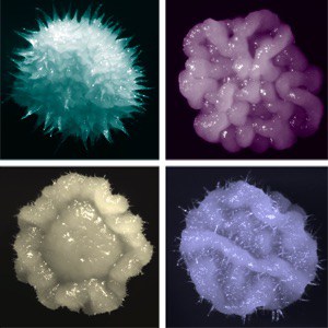
Candida albicans is an infectious fungus that is even scarier in action than in appearance. This funky fungus hides out in the body, waiting for the perfect moment to strike. When it senses that your immune system is weaker than usual, it attacks and causes disease.
Pictured here are some of the different creepy shapes Candida morphs into, which allow it to hide from your immune system and invade different tissues.
Submitted by PhD student Amanda Veri and postdoctoral fellow Teresa O’Meara, Cowen lab, University of Toronto.
LAKE OF SOULS
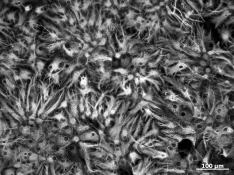
The cells in this picture are familiar to neuroscientists as ‘astrocytes,’ but their resemblance to trapped spirits floating in an eery lake just can’t be ignored. This could be a neuroscientist’s take on the River Styx, or the lake in the cave where Harry Potter and Dumbeldore search for the first horcrux in The Half-Blood Prince.
Submitted by Samantha Yammine, PhD student in the van der Kooy lab, University of Toronto.
THE EVER-WATCHFUL EYE
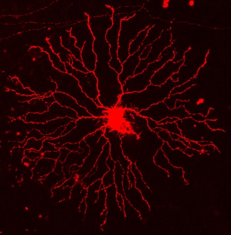
These bloodshot branches belong to a starburst amacrine cell, a type of neuron in the eye. Starburst cells are responsible for sensing the direction of moving targets. They use these eerie branches to communicate with neighbouring cells to transmit visual information to the brain.
Submitted by Samantha Esteves, MSc student in the Lefebvre lab at the Hospital for Sick Children.
THE MUTANTS ARE SPAWNING
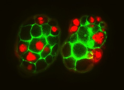
This picture is of Caenorhabditis elegans, which, although both it’s name and it’s appearance may imply extraterrestrial origin, is actually a transparent roundworm. Shown here are two embryos that have mutations in a gene important for cell division.
Submitted by Abigail Mateo, PhD candidate in the Derry lab at the Hospital for Sick Children.
THE BATTLE OF HELLS DEEP
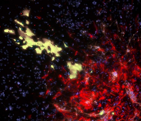
These ghost-like yellow cells, whose colouring is caused by a viral infection that makes them fluorescent, are plunging head first into the fiery depths of a cellular hell made of astrocytes.
Submitted by Samantha Yammine, PhD student in the Van Der Kooy lab at the University of Toronto.
RADIOACTIVE SPIDER WEB
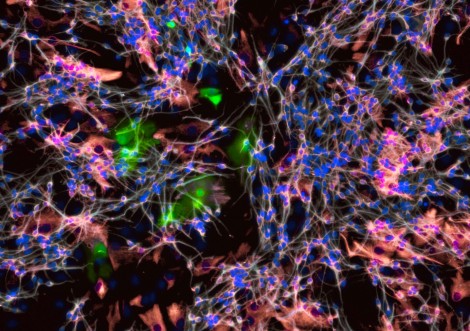
This web of cells is made up of neurons that were born from stem cells in a dish. They form complex connections with one another both in a dish and in the brain, and they often look like irregular spider webs when visualized. Some of the cells trapped in the web were infected with a virus.
Submitted by Samantha Yammine, PhD student in the van der Kooy lab at the University of Toronto.
DEEP, DEEP DREAMS
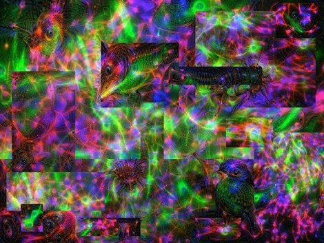
A representation of the confusing worlds we visit in our dreams. Created using the open source Google deep dream code, which reveals the psychedelic patterns hidden in the connections between brain cells.
Submitted by Samantha Yammine and Albi Celaj, PhD students in the van der Kooy and Roth lab (respectively) at the University of Toronto.
THE HAND THAT FEEDS
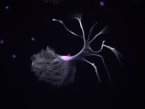
Not every cell made in a dish turns out perfectly, and this is an example of one that does not look quite as it should -— biologically speaking. It seems this cell just wanted to dress up for Halloween a little early!
Submitted by Samantha Yammine, PhD student in the van der Kooy lab, University of Toronto.