How can physicists extract information from mollusk shells, and how can we learn more about lung disease? Undergraduate women researchers explored these questions, and answered them at the Canadian Conference for Undergraduate Women in Physics (CCUWiP), which was hosted at the University of Toronto from January 17–19.
Kaitlyn Liang, one of the conference’s co-chairs, wrote to The Varsity that conferences such as CCUWiP “help female undergraduate physics majors through the creation of a community of independent and intelligent underrepresented minorities to further support and encourage each other throughout their personal and professional lives.”
CCUWiP hosted speakers, and held panels on physics and career paths and a variety of workshops, including an LGBTQ+ roundtable and one on combating racism. Liang hopes that “delegates take away a sense of community and pride in their identity as an underrepresented minority in physics.”
A highlight of the event was the presentation of research posters. The Varsity interviewed four students at the segment to learn more about their undergraduate research.
Analyzing the shells of mollusks
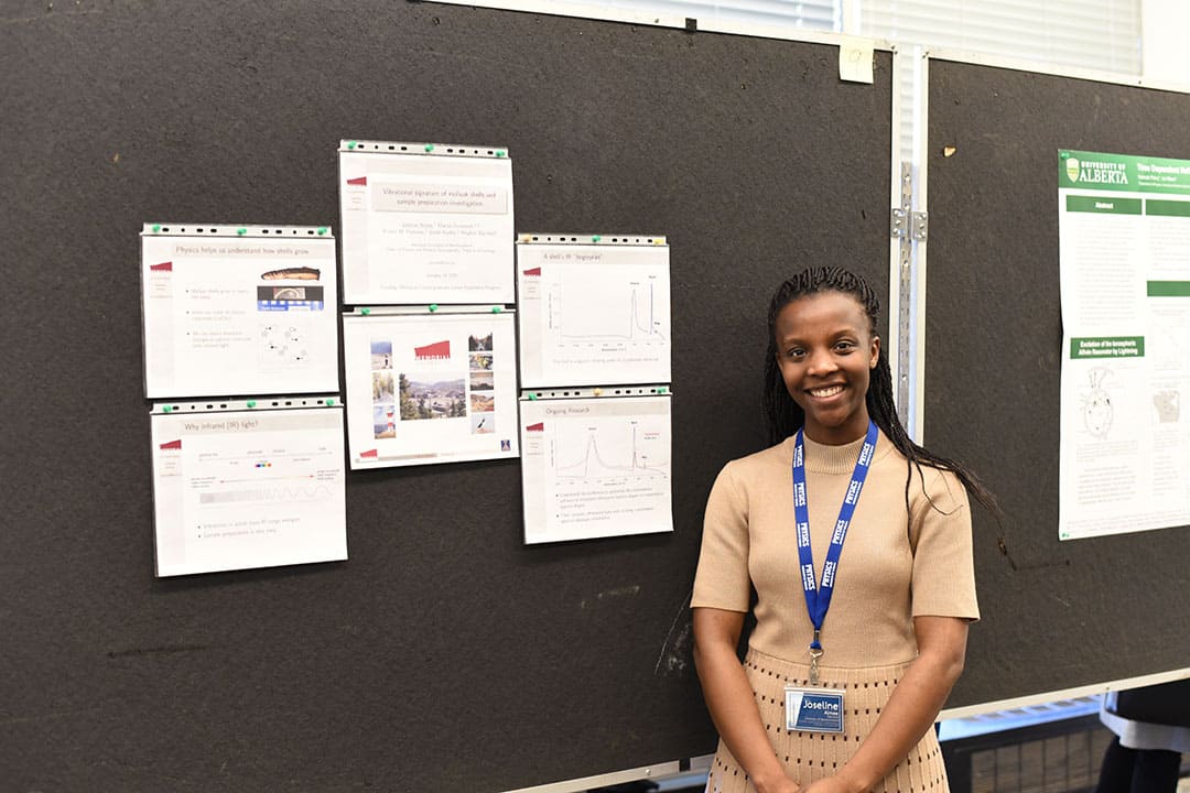
Joseline Aimee is a fourth-year student studying applied mathematics and physics at the Memorial University of Newfoundland. Her project was on the “vibrational signature of mollusk shells and sample preparation investigation,” she explained to The Varsity. For her project, she worked in conjunction with physicists and archaeologists in order “to understand how we can use infrared radiation to extract atomic level information of a material.”
The research team used both reflectance Fourier transform-infrared spectroscopy (FTIR) and transmission FTIR, which are analytical techniques, to examine the mollusk shells.
“For reflectance FTIR,” explained Aimee, “we just took a mollusk shell and a marine shell and we held it in front of a radiation source and we detected how much of that radiation [was] reflected by the material.”
The team analyzed the spectrum that resulted from the “interaction between the solid and the light [that was] detected.”
In contrast, transmission FTIR requires the sample to be grinded and diluted. “We’re using the same sample and we’re using the same infrared radiation source, but the slight change of the sample preparations gives us a different spectrum.”
“The main conclusion,” she explained, “is that further terms have to be considered to really understand the difference in these two spectras of the same material.”
Looking at different energy states
Chloe Cheng is a fourth-year U of T undergraduate studying astronomy and physics. Her research project, conducted in Vancouver over the summer, involved studying mercury.
Cheng studied the shape coexistence of the mercury-192 isotope, which she explained to The Varsity as a “phenomenon where the nucleus can exhibit excited states that are deformed.”
Her study involved the phenomenon of gamma decay. She explained that this occurs “when the nucleus decays from an excited energy state to a lower energy state by emitting something called a gamma ray.”
Her research also involved internal conversion, which occurs when “the gamma ray is still emitted from the nucleus, but it’s absorbed by one of the electrons orbiting the nucleus and its electron is emitted instead of the gamma rays,” she described.
Future steps for the study include further analysis of the shape coexistence in mercury-192.
Using beams for scans
Wajeeha Hassan is in her fourth year studying physics at the University of Calgary.
“The objective of this research,” said Hassan to The Varsity, “was to optimize the IDEAS beamline at the Canadian Light Source to get the best quality scans possible in order to continue to use the beamline in a way that would help us get the most accurate results over time.”
In order to accomplish this, they optimized both X-ray beamlines and the scan parameters. Next, the concentrations were tested to determine “how high or low [they] could go.” They concluded by doing linear combination fittings.
“Our main conclusion,” described Hassan, “[was] that our scans had been optimized so much so that we could be happy with our linear combination fitting.”
Studying biological structures
Ashlyn McNabb, a fourth-year undergraduate student studying medical physics at Ryerson University, looked at chronic obstructive pulmonary disease (COPD), more specifically, at the spatial interrelationship between small airways disease and emphysema.
McNabb explained that she re-oriented the CT images of lungs and looked at them using five different metrics.
She noted that here research data supports that emphysema is connected to “small airways disease, whereas small airways disease isn’t always connected to emphysema.”