
Arthritis and other joint disorders are among the top causes of disabilities in North America today. Joint pain is most common among people over the age of 50, but also affects many young people who suffer injuries from high impact sports. Researchers from the University of Toronto are using 3D printing technology to revolutionize treatment with biological joint replacements, a promising alternative to the artificial joint replacement surgeries that have become routine procedures.
Osteoarthritis, the breakdown of cartilage between bones, is the most common type of chronic joint disorder. Approximately one in six Canadians aged 15 years and older report symptoms of arthritis, yet surgery for joint replacement is usually the last line of treatment, often delayed for as long as possible. Currently, artificial joints are synthesized from metals and plastics that break down after 10 to 20 years and can erode or loosen even sooner. These artificial replacements may not provide a long-term solution for people with joint disorders.
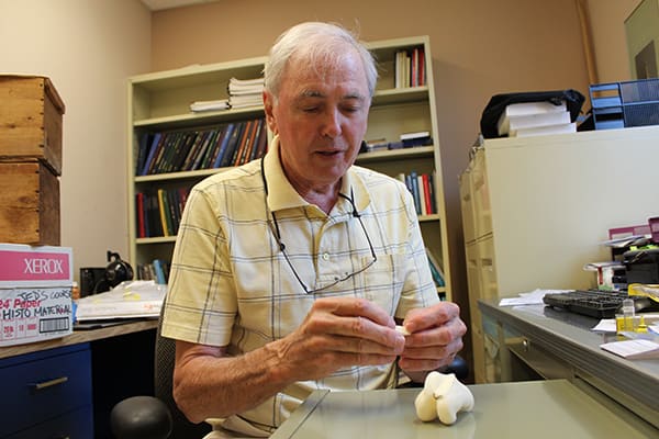
Dr. Robert Pilliar of the IBBME exhibits the template bone model. MEDIA PHOTO
Scientists at the University of Toronto are now taking part in a multidisciplinary collaborative project with partners from other Ontario universities including Waterloo, Guelph, McMaster, and Queens. The project aims to create biological joint replacements using 3D printing technology to provide longer-lasting joints that are catered to the individual.
Dr. Rita Kandel, chief of pathology at Mount Sinai Hospital and director of the Collaborative Program in Musculoskeletal Sciences at U of T, is part of the team that heads this research.
“Currently, if you damage your joint, there is no good way to repair it,” says Kandel. “Metal and plastic doesn’t belong in the body and cannot repair itself. By generating a biological implant using the individual’s own cells, it will reconstruct a normal, native joint surface,” she adds.
These biological joint replacements will overcome the many deficits of available technology. Cartilage — the soft connective tissue that is fused to bone — does not typically self-repair, but the biological joint replacement helps the body do just that.
The process begins by taking X-ray images of the damaged joint. Using these images, a 3D printer deposits porous material at certain locations to reconstruct the bone shape in the form of a bone substitute. This porous bone substitute matches the bone shape and size of each individual and is able to support the growth of cells and formation of cartilage. By integrating cartilage to the top surface of the bone substitute, bone can grow into the porous substitute. The bone substitute eventually degrades, leaving the whole area replaced with the patient’s own bone and cartilage. “It’s the part I call ‘personalized’ to the individual,” says Kandel.
The team is currently working toward the implementation of the procedure. Kandel estimates that clinical trials will be a possibility in as soon as five years.
“I believe it’s the way of the future,” says Kandel.